What is heart rhythm?
Heart Rhythm represents the place of origin of the electrical activity of the heart. In a normal healthy heart, electrical activity is started from the sinoatrial node, located on the upper part of the right atrium wall. In a normal healthy heart, electrical activity is generated and transmitted to the lower part of the Atrium. Then it stimulates the AV node. Then the electrical activity or impulses are sent via his bundle. Then this bundle divides into two branches. They are the right bundle and left bundle. Right bundle supplies to the right side of the heart, including the right side ventricular wall and their part of the septum. The left branch supplies the left ventricular wall and part of the septum. These cells are specified cardiac muscle cells. But this type of cell can repolarize and depolarize.
There are four types of cardiac rhythms.
- Sinus rhythm
- Atrial rhythm
- Nodal rhythm
- Ventricular Rhythm
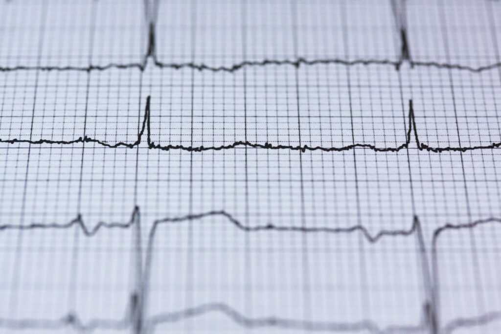
Sinus rhythm
Sinus rhythm is the normal cardiac rhythm of a healthy person. Sinus rhythm starts from the SA node. In ECG, atrial contraction is represented by the P wave of the ECG. In ECG, all p waves are followed by the QRS complexes meaning sinus rhythm is present.
Atrial Rhythm
Atrial rhythm means the heart’s electrical activity is driven by somewhere in the Atrium except the SA node. If it is the SA node, it is the sinus rhythm. Here the electrical pathway is abnormal in the atria. But below the AV node, it is normal. That means the QRS complex is intact. There are two common types of atrial rhythms.
- Atrial Fibrillation
- Atrial Flutter
Atrial Fibrillation: In atrial fibrillation, electrical activity starts from the right Atrium but in several places. In ECG, p waves can’t see clearly, and there is an irregular small wave pattern instead. QRS complexes are regular but irregular.
Atrial Flutter: In here, ECG has seen tooth appearance. Normal QRS complexes with regular or irregular patterns. The electrical activity starts from the right Atrium. But the electrical impulse is circulated inside the muscle wall of the Atrium. Because of this, it generates a saw tooth like the same pattern repeatedly.
Nodal rhythm
Here electrical activity starts from the atrioventricular (AV) node. But the pathway beyond that is normal. That means the QRS complex is intact. But atria depolarize in the reverse direction. In ECG, retrograde p waves can be seen. But in practice, it is challenging to see the retrograde p waves that it can also take as absent p waves.
Ventricular Rhythm
The ventricles drive electrical activity. This is the only broad category with abnormal QRS complexes. In ECG, broad QRS complexes are common. And the rate is high. According to that, there are two types of ventricular rhythms.
- Monomorphic ventricular tachycardia
- Polymorphic ventricular tachycardia
Tachycardia means heart rate is higher than 100 beats per minute. Monomorphic means the wave pattern is the same. The height of the waves is the same. Polymorphic have variable sizes and different types of locks.